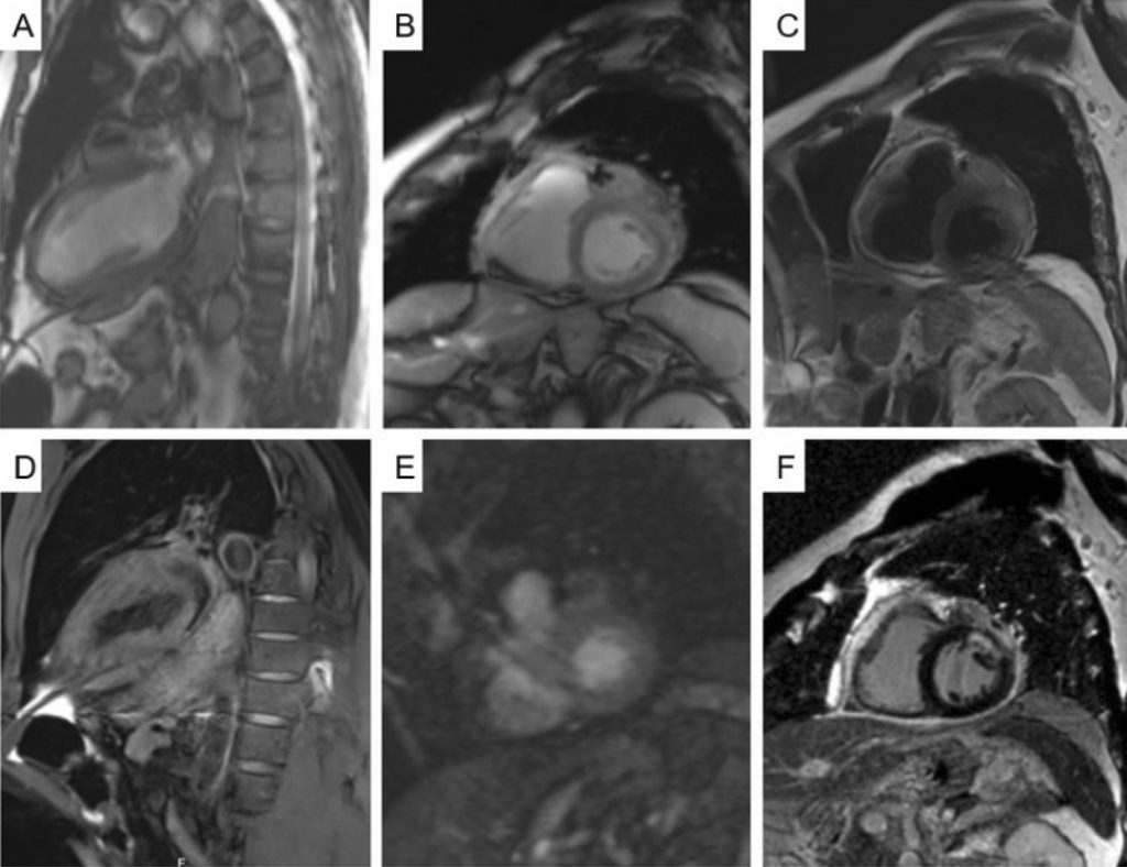
Metastases to the heart and pericardium are much more common than primary cardiac tumors and are generally associated with a poor prognosis. , While they are most commonly asymptomatic, cardiac metastases can mimic primary cardiac diseases such as acute coronary syndromes, congestive heart failure and pericarditis. , Lung cancer is the most frequent source of metastatic cardiac disease, either from direct extension or by a combination of lymphatic, hematogenous, and transvenous dissemination. ,
[…]
A Case of Acute Myocardial Infarction and Pericarditis Unmasking Metastatic Involvement of the Heart
TorresS, VasconcelosM, SousaC, MadureiraAJ, NunesA, MacielMJ. A Case of Acute Myocardial Infarction and Pericarditis Unmasking Metastatic Involvement of the Heart. Arq. Bras. Cardiol. 2020;115(1 suppl 1):22-4.
Torres,Sofia; Vasconcelos,Mariana; Sousa,Carla; Madureira,Antonio J.; Nunes,Alzira; Maciel,Maria Júlia. A Case of Acute Myocardial Infarction and Pericarditis Unmasking Metastatic Involvement of the Heart. Arq. Bras. Cardiol., v. 115, n. 1 suppl 1, p. 22-24, Sep. 2020.
Torres,S., Vasconcelos,M., Sousa,C., Madureira,A.J., Nunes,A., & Maciel,M.J. (2020). A Case of Acute Myocardial Infarction and Pericarditis Unmasking Metastatic Involvement of the Heart. Arq. Bras. Cardiol.,115(1 suppl 1), 22-24.
Torres,Sofia and Vasconcelos,Mariana and Sousa,Carla and Madureira,Antonio J. and Nunes,Alzira and Maciel,Maria Júlia. A Case of Acute Myocardial Infarction and Pericarditis Unmasking Metastatic Involvement of the Heart. Arq. Bras. Cardiol. [online]. 2020, vol. 115, n. 1 suppl 1, [cited 2025-09-07], pp.22-24. Available from: <https://abccardiol.org/en/article/a-case-of-acute-myocardial-infarction-and-pericarditis-unmasking-metastatic-involvement-of-the-heart/>. ISSN 0066-782X.
Figure 1
– cMRI (A) (B) b-SSFP cine images revealing an elongated mass (measuring 25 x 13 x 40 mm) adjacent to the basal anterior and antero-lateral segments and in close contact with the LAD artery stent. (C) High intensity signal on T2-weighted images. (D) Isointense signal intensity on T1-weighted images. (E) First pass perfusion of the mass. (F) LGE with heterogeneous appearance of the mass and diffuse contrast-enhanced pericardium.