Arq. Bras. Cardiol. 2023; 120(7): e20220762
Cardiac Magnetic Resonance Imaging in a 7 Tesla Magnetic Field: Initial Experience with Hydrogen and Sodium Nuclei
Carlos E. Rochitte
![]() , Douglas C. Silva, Maria C. Otaduy, Khallil T. Chaim, Cesar H. Nomura, Bruno Caramelli
, Douglas C. Silva, Maria C. Otaduy, Khallil T. Chaim, Cesar H. Nomura, Bruno Caramelli
Introduction
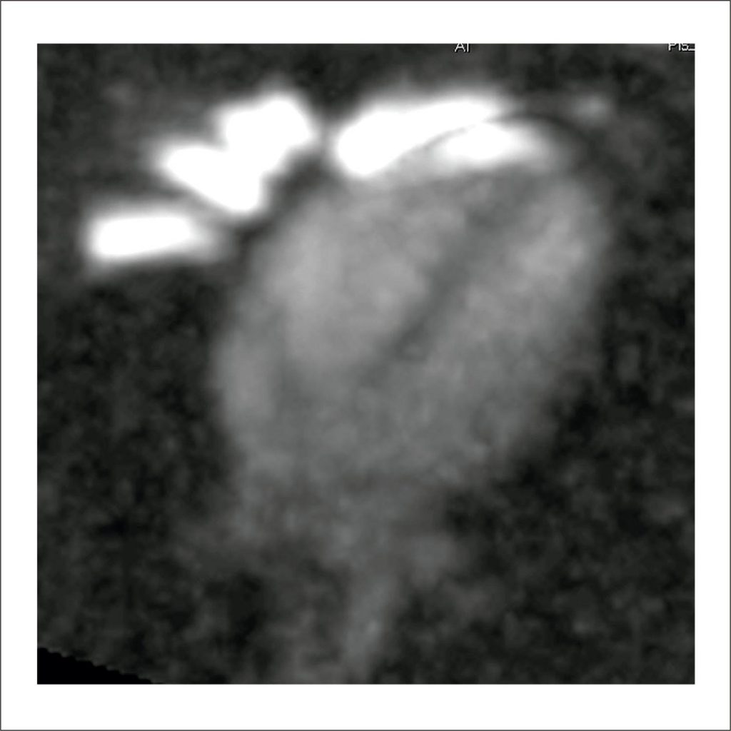
The cellular integrity of myocardial tissue is crucial information to evaluate the viability of the heart. Cardiac magnetic resonance imaging (CMR) is considered the gold standard method for functional analysis of the heart, and it plays an essential role in the diagnosis of several cardiomyopathies, including myocardial infarction. When there is a failure in the energy supply of heart cells, there occurs a decrease in blood flow in certain regions, which can lead to cell death. CMR can assess the viability of cardiac tissue by means of interaction with the protons present in this tissue, the hydrogen nucleus being the most common for this purpose.
In recent years, another nucleus has emerged as a possible ischemic marker of the heart, namely, sodium. Studies carried out in animals have shown that the evolution of the ischemic process is directly correlated with the accumulation of intracellular sodium. In this ischemic process, the function of the sodium-potassium pump is compromised, generating an imbalance in the concentration of this electrolyte, , , leading to influx of sodium into the intracellular environment. Studies also suggest that sodium concentration is directly related to the extent of ischemic injury. Sodium concentration is higher in the extracellular environment in normal physiological situations, but with failure in the function of the sodium-potassium pump, there is an influx of sodium into the intracellular environment, thus making it a potential marker for ischemic events in the heart. , , ,
[…]
886