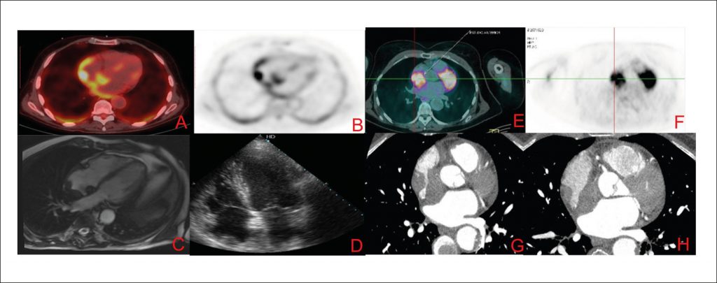
Erdheim-Chester Disease is a rare entity, classified as an inflammatory myeloid neoplasm, with an unknown incidence, occurring preferentially in men after 50 years of age. Classically, it has a multisystemic presentation, with the skeletal system being the most frequently affected (90% of the patients), followed by genitourinary involvement in 60% of cases and central nervous system in the pituitary and diabetes insipidus in 25% of the cases. Cardiovascular manifestations are present in more than half of the patients, with aortic infiltration and atrial pseudotumor being the most common forms.
Cardiovascular Manifestations of Erdheim-Chester’s Disease: A Case
Series
CostaIBSS, AbdoANR, BittarCS, FonsecaSMR, MoraesASHT, Kalil FilhoR, PereiraJ, et al. Cardiovascular Manifestations of Erdheim-Chester’s Disease: A Case
Series. Arq. Bras.
Cardiol. 2018;111(6):852-5.
Costa,Isabela Bispo Santos da Silva; Abdo,André Neder Ramires; Bittar,Cristina Salvadori; Fonseca,Silvia Moulin Ribeiro; Moraes,Aline Sabrina Holanda Teixeira; Kalil Filho,Roberto; Pereira,Juliana; Hajjar,Ludhmila Abrahão. Cardiovascular Manifestations of Erdheim-Chester’s Disease: A Case
Series. Arq. Bras.
Cardiol., v. 111, n. 6, p. 852-855, Dec. 2018.
Costa,I.B.S.S., Abdo,A.N.R., Bittar,C.S., Fonseca,S.M.R., Moraes,A.S.H.T., Kalil Filho,R., Pereira,J., & Hajjar,L.A. (2018). Cardiovascular Manifestations of Erdheim-Chester’s Disease: A Case
Series. Arq. Bras.
Cardiol.,111(6), 852-855.
Costa,Isabela Bispo Santos da Silva and Abdo,André Neder Ramires and Bittar,Cristina Salvadori and Fonseca,Silvia Moulin Ribeiro and Moraes,Aline Sabrina Holanda Teixeira and Kalil Filho,Roberto and Pereira,Juliana and Hajjar,Ludhmila Abrahão. Cardiovascular Manifestations of Erdheim-Chester’s Disease: A Case
Series. Arq. Bras.
Cardiol. [online]. 2018, vol. 111, n. 6, [cited 2025-10-24], pp.852-855. Available from: <https://abccardiol.org/en/article/cardiovascular-manifestations-of-erdheim-chesters-disease-a-caseseries/>. ISSN 0066-782X.
Figure 1
Images A to D refer to case 1 and the images from E to H refer to
case 2. Images A and B represent images of 18-FDG PET-CT showing
lesion in the right atrium roof. The C image represent CMR image,
SSFP cine 4 chambers with hypointense lesion in the right atrium
roof. The D image represents a transthoracic echocardiogram image
with the same topography. The images E and F represent 18-FDG PET-CT
with capturing lesion in the right atrium and G and H images
represent contrast computed tomography showing evidence of expansive
right atrial.s