Case 3/2020 – Pulmonary Atresia, Interventricular Communication and Anomalous Origin of the Right Pulmonary Artery from the Ascending Aorta developing after Prior Left Central Shunt, in a Symptomatic 40-year-old Adult.
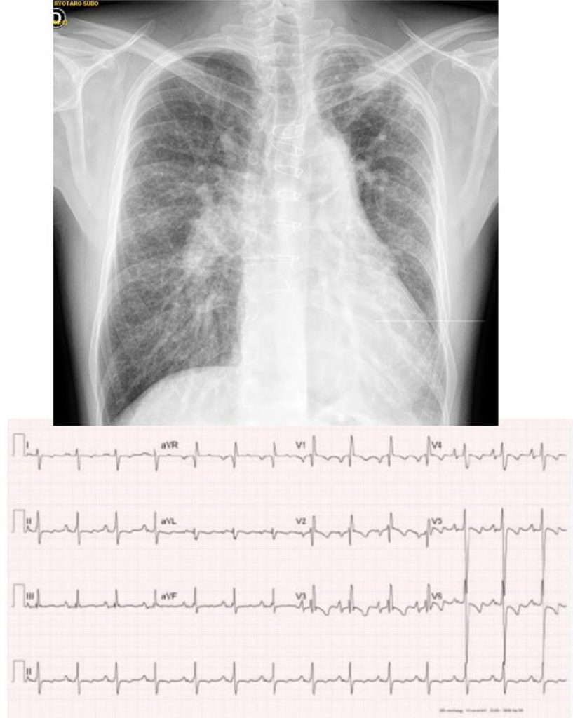
Electrocardiogram: Sinus rhythm, signs of right cavity overload, with apiculate P wave + 70º, QRS complex showing predominance of S waves from V4 to V6 and axis deviated to the right (AQRS= +110°). The T wave was negative in the precordial leads with diffuse ventricular repolarization changes in all other leads ( ).
[…]
Case 3/2020 – Pulmonary Atresia, Interventricular Communication and Anomalous Origin of the Right Pulmonary Artery from the Ascending Aorta developing after Prior Left Central Shunt, in a Symptomatic 40-year-old Adult.
AtikE, BinottoMA, BarretoAC. Case 3/2020 – Pulmonary Atresia, Interventricular Communication and Anomalous Origin of the Right Pulmonary Artery from the Ascending Aorta developing after Prior Left Central Shunt, in a Symptomatic 40-year-old Adult.. Arq. Bras. Cardiol. 2020;114(4 suppl 1):43-6.
Atik,Edmar; Binotto,Maria Angélica; Barreto,Alessandra Costa. Case 3/2020 – Pulmonary Atresia, Interventricular Communication and Anomalous Origin of the Right Pulmonary Artery from the Ascending Aorta developing after Prior Left Central Shunt, in a Symptomatic 40-year-old Adult.. Arq. Bras. Cardiol., v. 114, n. 4 suppl 1, p. 43-46, May. 2020.
Atik,E., Binotto,M.A., & Barreto,A.C. (2020). Case 3/2020 – Pulmonary Atresia, Interventricular Communication and Anomalous Origin of the Right Pulmonary Artery from the Ascending Aorta developing after Prior Left Central Shunt, in a Symptomatic 40-year-old Adult.. Arq. Bras. Cardiol.,114(4 suppl 1), 43-46.
Atik,Edmar and Binotto,Maria Angélica and Barreto,Alessandra Costa. Case 3/2020 – Pulmonary Atresia, Interventricular Communication and Anomalous Origin of the Right Pulmonary Artery from the Ascending Aorta developing after Prior Left Central Shunt, in a Symptomatic 40-year-old Adult.. Arq. Bras. Cardiol. [online]. 2020, vol. 114, n. 4 suppl 1, [cited 2025-10-24], pp.43-46. Available from: <https://abccardiol.org/en/article/case-3-2020-pulmonary-atresia-interventricular-communication-and-anomalous-origin-of-the-right-pulmonary-artery-from-the-ascending-aorta-developing-after-prior-left-central-shunt-in-a-symp/>. ISSN 0066-782X.
Figure 1
– Chest X-ray showing enlarged cardiac area with left ventricular dominance and increased pulmonary vascular network in the right hila, with a reduction towards the lower lobe. It is reduced to the left with thinner blood vessels. Electrocardiogram shows right cavity overload and diffuse ventricular repolarization changes.