Arq. Bras. Cardiol. 2023; 120(11): e20220822
Coronary Inflammation by Computed Tomography Pericoronary Fat Attenuation and Increased Cytokines in Young Male Anabolic Androgenic Steroid Users
Francis Ribeiro de Souza
![]() , Carlos E. Rochitte
, Carlos E. Rochitte
![]() , Douglas Carli Silva
, Douglas Carli Silva
![]() , Barbara Sampaio, Marisa Passarelli, Marcelo R. dos Santos, Guilherme W. Fonseca, Antonio Carlos Battaglia Filho, Kelly Correa, Renata Margarida do Val, Maurício Yonamine, Rosa Maria R. Pereira, Carlos Eduardo Negrão, Roberto Kalil-Filho, Maria Janieire de Nazaré Nunes Alves
, Barbara Sampaio, Marisa Passarelli, Marcelo R. dos Santos, Guilherme W. Fonseca, Antonio Carlos Battaglia Filho, Kelly Correa, Renata Margarida do Val, Maurício Yonamine, Rosa Maria R. Pereira, Carlos Eduardo Negrão, Roberto Kalil-Filho, Maria Janieire de Nazaré Nunes Alves
This Original Article is referred by the Short Editorial "Pericoronary Fat Attenuation on Computed Tomography Unveils a Guilty Factor of Coronary Artery Disease Associated with Anabolic–Androgenic Steroids".
Abstract
Background
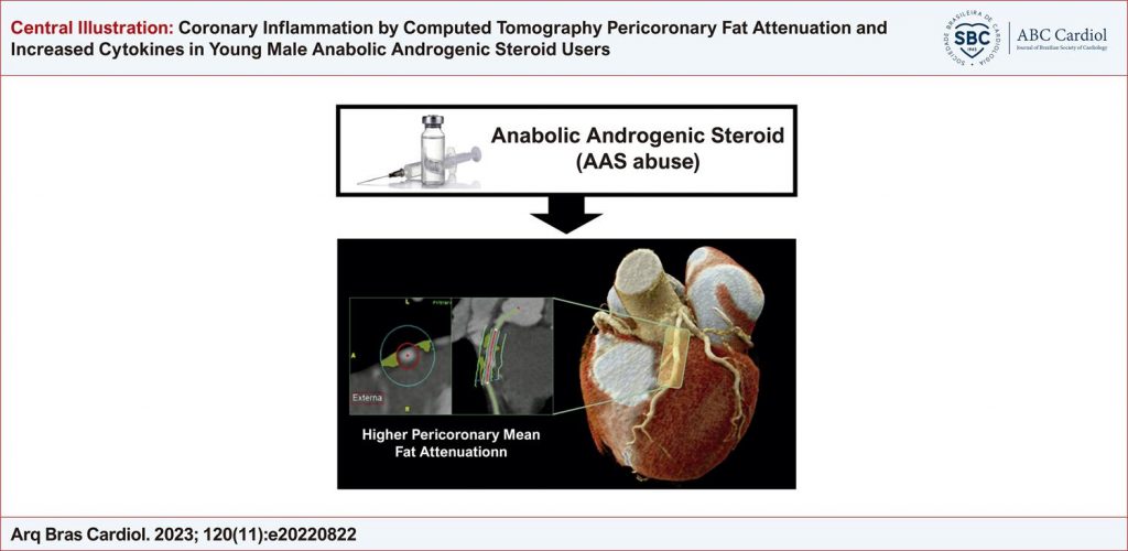
Anabolic androgenic steroid (AAS) abuse has been associated with coronary artery disease (CAD). Pericoronary fat attenuation (pFA) is a marker of coronary inflammation, which is key in the atherosclerotic process.
Objective
To evaluate pFA and inflammatory profile in AAS users.
Methods
Twenty strength-trained AAS users (AASU), 20 AAS nonusers (AASNU), and 10 sedentary controls (SC) were evaluated. Coronary inflammation was evaluated by mean pericoronary fat attenuation (mPFA) in the right coronary artery (RCA), left anterior descending coronary artery (LAD), and left circumflex (LCx). Interleukin (IL)-1 (IL-1), IL-6, IL-10, and TNF-alpha were evaluated by optical density (OD) in a spectrophotometer with a 450 nm filter. P<0.05 indicated statistical significance.
Results
AASU had higher mPFA in the RCA (-65.87 [70.51-60.70] vs. -78.07 [83.66-72.87] vs.-78.46 [85.41-71.99] Hounsfield Units (HU), respectively, p<0.001) and mPFA in the LAD (-71.47 [76.40-66.61] vs. -79.32 [84.37-74.59] vs. -82.52 [88.44-75.81] HU, respectively, p=0.006) compared with AASNU and SC. mPFA in the LCx was not different between AASU, AASNU, and SC (-72.41 [77.17-70.37] vs. -80.13 [86.22-72.23] vs. -78.29 [80.63-72.29] HU, respectively, p=0.163). AASU compared with AASNU and SC, had higher IL-1, (0.975 [0.847-1.250] vs. 0.437 [0.311-0.565] vs. 0.530 [0.402-0.780] OD, respectively, p=0.002), IL-6 (1.195 [0.947-1.405] vs. 0.427 [0.377-0.577] vs. 0.605 [0.332-0.950] OD, p=0.005) and IL-10 (1.145 [0.920-1.292] vs. 0.477 [0.382-0.591] vs. 0.340 [0.316-0.560] OD, p<0.001). TNF-α was not different between the AASU, AASNU, and SC groups (0.520 [0.250-0.610] vs. 0.377 [0.261-0.548] vs. 0.350 [0.182-430]), respectively.
Conclusion
Compared with ASSNU and controls, AASU have higher mPFA and higher systemic inflammatory cytokines profile suggesting that AAS may induce coronary atherosclerosis through coronary and systemic inflammation.
1,527