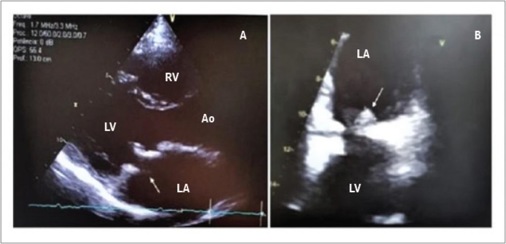
Coxiella burnetii infective endocarditis (IE) represents a rarely reported zoonosis in Brazil. It is estimated that Coxiella burnetii is responsible for up to 5% of all IE cases worldwide. The disease affects mostly valvulopathy patients and immunocompromised subjects.
Different from the classical acute and sub-acute endocarditis, the clinical picture is frustrating, and, because this is an obligate intracellular microorganism, hemocultures (HMC) are predominantly negative, which makes the clinical suspicion more difficult.
[…]
Coxiella Burnetii Endocarditis: Can Positron Emission Tomography be an Alternative to Diagnosis?
MizutaMH, RomeroCE, VintimillaSC, LealTCAT, SoaresPR, SoeiroAM. Coxiella Burnetii Endocarditis: Can Positron Emission Tomography be an Alternative to Diagnosis?. Arq. Bras. Cardiol. 2022;118(6):1144-6.
Mizuta,Marjorie Hayashida; Romero,Cristhian Espinoza; Vintimilla,Santiago Castro; Leal,Tatiana de Carvalho Andreucci Torres; Soares,Paulo Rogério; Soeiro,Alexandre de Matos. Coxiella Burnetii Endocarditis: Can Positron Emission Tomography be an Alternative to Diagnosis?. Arq. Bras. Cardiol., v. 118, n. 6, p. 1144-1146, Jun. 2022.
Mizuta,M.H., Romero,C.E., Vintimilla,S.C., Leal,T.C.A.T., Soares,P.R., & Soeiro,A.M. (2022). Coxiella Burnetii Endocarditis: Can Positron Emission Tomography be an Alternative to Diagnosis?. Arq. Bras. Cardiol.,118(6), 1144-1146.
Mizuta,Marjorie Hayashida and Romero,Cristhian Espinoza and Vintimilla,Santiago Castro and Leal,Tatiana de Carvalho Andreucci Torres and Soares,Paulo Rogério and Soeiro,Alexandre de Matos. Coxiella Burnetii Endocarditis: Can Positron Emission Tomography be an Alternative to Diagnosis?. Arq. Bras. Cardiol. [online]. 2022, vol. 118, n. 6, [cited 2025-10-23], pp.1144-1146. Available from: <https://abccardiol.org/en/article/coxiella-burnetii-endocarditis-can-positron-emission-tomography-be-an-alternative-to-diagnosis/>. ISSN 0066-782X.
Figure 1
A) 2D TTE in longitudinal parasternal position showing mitral prosthesis with thickened leaflets and pannus aspect. B) 2D TEE at 60º showing an oval shape adhered to the posterior leaflet atrial face. RV: right ventricle; LV: left ventricle; AO: aorta; LA: left atrium.