Arq. Bras. Cardiol. 2024; 121(6): e20230442
Echocardiographic Alterations of Cardiac Geometry and Function in Patients with Familial Partial Lipodystrophy
Minna Moreira Dias Romano
![]() , André Timóteo Sapalo, Natália Rossin Guidorizzi
, André Timóteo Sapalo, Natália Rossin Guidorizzi
![]() , Henrique Turin Moreira
, Henrique Turin Moreira
![]() , Paula Ananda Chacon Inês
, Paula Ananda Chacon Inês
![]() , Lucas Candelária Kalil, Maria Cristina Foss, Francisco José Albuquerque de Paula
, Lucas Candelária Kalil, Maria Cristina Foss, Francisco José Albuquerque de Paula
This Original Article is referred by the Short Editorial "Understanding Cardiac Alterations in Familial Partial Lipodystrophy: Insights from Echocardiography".
Abstract
Background:
Cardiomyopathy associated with partial lipodystrophy (PL) has not been well described yet.
Objective:
To characterize cardiac morphology and function in PL.
Methods:
Patients with familial PL and controls were prospectively assessed by transthoracic echocardiography and with speckle-tracking echocardiography (global longitudinal strain, GLS). The relationship between echocardiographic variables and PL diagnosis was tested with regression models, considering the effect of systolic blood pressure (SBP). Significance level of 5% was adopted.
Results:
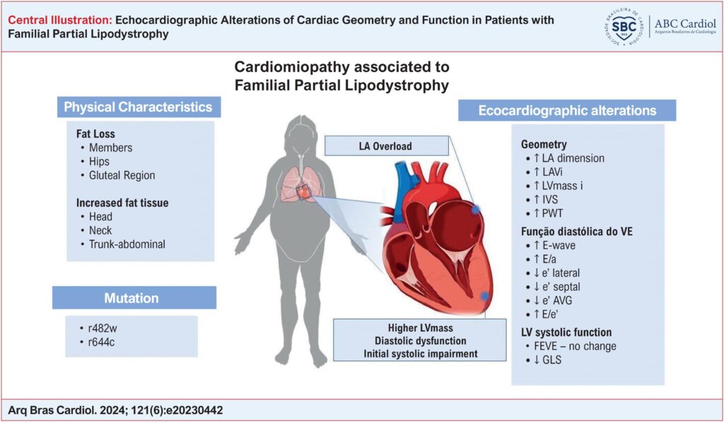
Twenty-nine patients with PL were compared to 17 controls. They did not differ in age (p=0.94), gender or body mass index (p= 0.05). Patients with PL had statistically higher SBP (p=0.02) than controls. Also, PL patients had higher left atrial dimension (37.3 ± 4.4 vs. 32.1 ± 4.3 mm, p= 0.001) and left atrial (30.2 ± 7.2 vs. 24.9 ± 9.0 mL/m2,p=0.02), left ventricular (LV) mass (79.3 ± 17.4 vs. 67.1 ± 19.4, p=0.02), and reduced diastolic LV parameters (E’ lateral, p= 0.001) (E’ septal, p= 0.001), (E/E’ ratio, p= 0.02). LV ejection fraction (64.7 ± 4.6 vs. 62.2 ± 4.4 %, p= 0.08) and GLS were not statistically different between groups (-17.1 ± 2.7 vs. -18.0 ± 2.0 %, p= 0.25). There was a positive relationship of left atrium (β 5.6, p<0.001), posterior wall thickness, (β 1.3, p=0.011), E’ lateral (β -3.5, p=0.002) and E’ septal (β -3.2, p<0.001) with PL diagnosis, even after adjusted for SBP.
Conclusion:
LP patients have LV hypertrophy, left atrial enlargement, and LV diastolic dysfunction although preserved LVEF and GLS. Echocardiographic parameters are related to PL diagnosis independent of SBP.
Keywords: Echocardiography; Heart Function Tests; Lipodystrophy
882