Arq. Bras. Cardiol. 2019; 112(6): 803-806
Extensive Anterior Myocardial Infarction … and Something Else?
Andrés Ricardo Pérez Riera
![]() , Raimundo Barbosa Barros
, Raimundo Barbosa Barros
![]() , Antônio Fernandes Silva e Sousa Neto
, Antônio Fernandes Silva e Sousa Neto
![]() , Rodrigo Daminello Raimundo
, Rodrigo Daminello Raimundo
![]() , Luiz Carlos de Abreu
, Luiz Carlos de Abreu
![]() , Kjell Nikus
, Kjell Nikus
![]()
DOI: 10.5935/abc.20190096
Case report
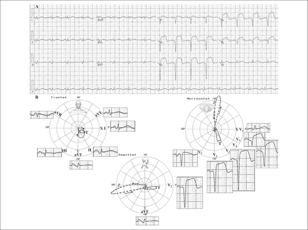
A 64-year-old Caucasian female, with a one-week history of stress angina. She was admitted to the hospital 2 hours after the onset of oppressive retrosternal pain at rest.
Risk factors: hypertension, smoker, dyslipidemia and diabetes.
[…]
Extensive Anterior Myocardial Infarction … and Something
Else?
1,050