Arq. Bras. Cardiol. 2017; 108(4): 381-382
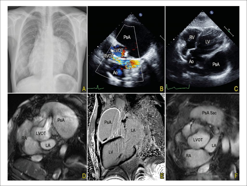
Giant Left Ventricle Outflow Tract Pseudoaneurysm after Ross Procedure
DOI: 10.5935/abc.20170008
A 33-year-old woman was admitted to our hospital because of dyspnea on exertion, orthopnea, cough and pedal edema for the past six months. Six years earlier she had been submitted to Ross procedure for correction of a bicuspid aortic valve.
Physical exam was unremarkable except by a grade 3 systolic murmur on left sternal border.
[…]
Giant Left Ventricle Outflow Tract Pseudoaneurysm after Ross
Procedure
283