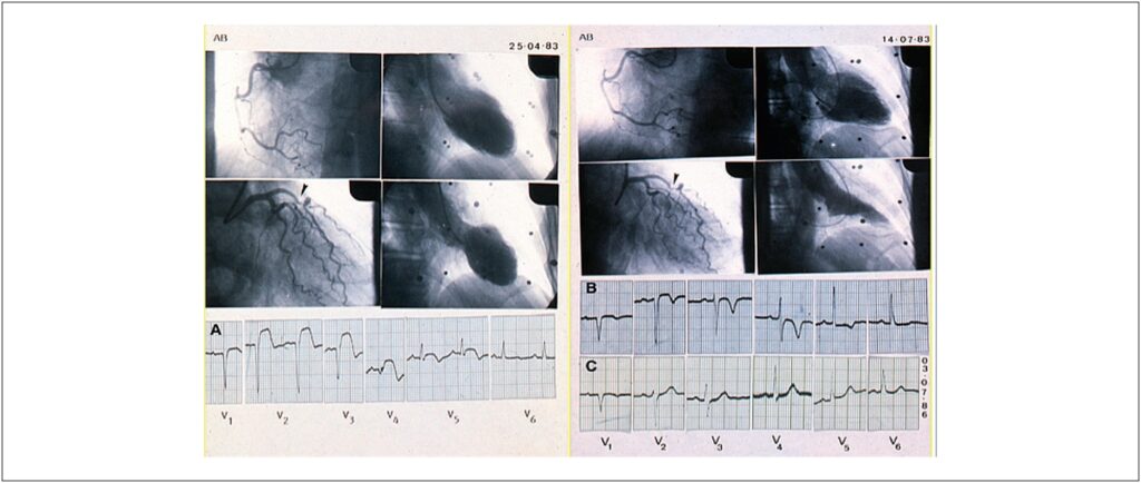
Plaque rupture and erosion are the most common immediate pathologies associated with acute myocardial infarction. However, other mechanisms must be considered. The reported case occurred at a time when percutaneous or pharmacological coronary revascularization were not the standard treatment, which sparked discussion about these other mechanisms, as well as the possible advantages associated with the myocardial revascularization procedures.
[…]
Interpreting Acute Myocardial Infarction 40 Years Later. Evolution of Knowledge: What is the Best Explanation?
AndradeVC, RezendePC, HuebW, RamiresJAF. Interpreting Acute Myocardial Infarction 40 Years Later. Evolution of Knowledge: What is the Best Explanation?. Arq. Bras. Cardiol. 2024;121(3):e20230757.
Andrade,Vitor Coutinho; Rezende,Paulo Cury; Hueb,Whady; Ramires,José Antonio Franchini. Interpreting Acute Myocardial Infarction 40 Years Later. Evolution of Knowledge: What is the Best Explanation?. Arq. Bras. Cardiol., v. 121, n. 3, e20230757, Apr. 2024.
Andrade,V.C., Rezende,P.C., Hueb,W., & Ramires,J.A.F. (2024). Interpreting Acute Myocardial Infarction 40 Years Later. Evolution of Knowledge: What is the Best Explanation?. Arq. Bras. Cardiol.,121(3), e20230757.
Andrade,Vitor Coutinho and Rezende,Paulo Cury and Hueb,Whady and Ramires,José Antonio Franchini. Interpreting Acute Myocardial Infarction 40 Years Later. Evolution of Knowledge: What is the Best Explanation?. Arq. Bras. Cardiol. [online]. 2024, vol. 121, n. 3, [cited 2025-10-22], e20230757. Available from: <https://abccardiol.org/en/article/interpreting-acute-myocardial-infarction-40-years-later-evolution-of-knowledge-what-is-the-best-explanation/>. ISSN 0066-782X.
Figure 1
Immediate and late coronary cineangiography and ECG images. CAPTION: Upper left frame: Sub occlusion of the anterior descending artery. Circumflex and right coronary arteries: absence of obstructions. Left ventricle: severe systolic dysfunction. Upper right frame (three months later). Similar angiogram with recovery of the left ventricle function. ECG (A): ST-segment elevation in precordial leads. ECG (B): ST-segment recovery and T wave inversion only. ECG (C): Three years later: Close to normal.