Arq. Bras. Cardiol. 2024; 121(6): e20230732
Multiple Intracardiac Metastases – A Chaplet Heart
Mariana Tinoco
![]() , Margarida Castro
, Margarida Castro
![]() , Hans Dabó, Filipa Cordeiro, Pedro von Hafe, António Lourenço
, Hans Dabó, Filipa Cordeiro, Pedro von Hafe, António Lourenço
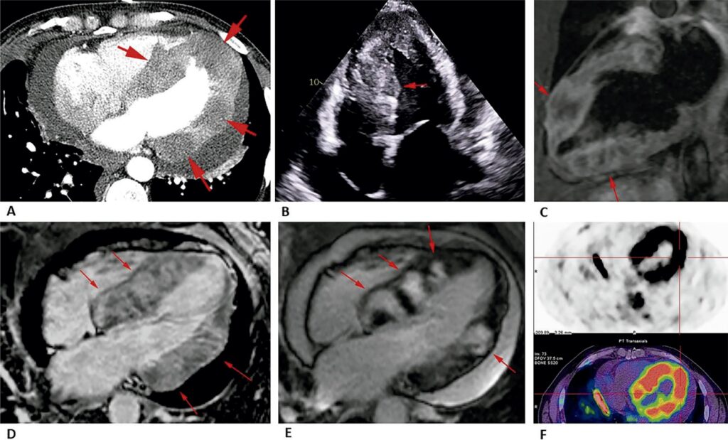
A 52-year-old male was undergoing first-line chemotherapy with pemetrexed due to a stage IV right hilar lung adenocarcinoma with likely pleural, pericardial, liver, and bone metastases. During follow-up, no evidence of disease progression was found. Four years later a follow-up chest CT scan documented a nodular heterogeneous enhancement of the left ventricular (LV) myocardium (), the remaining lesions were overlapping. He had no cardiovascular symptoms and clinical examination was unremarkable. Transthoracic echocardiography () and cardiac MRI (Figures 1C-E) revealed several well-rounded endo-myocardial masses, some of which multilobulated sometimes with almost transmural extension. The lesions are predominantly distributed along the LV inferoseptal wall, inferolateral wall, anterolateral wall, anterior wall, and right side of the interventricular septum. These lesions had heterogeneous signal characteristics, being predominantly hyperintense on T1-weighted images, hypointense on T2-weighted images with a peripheral halo of hyperintensity, showing early heterogeneous and late intense gadolinium enhancement; and abnormal uptake of fluorodeoxyglucose in a 18F-FDG PET/CT ().
[…]
384