Arq. Bras. Cardiol. 2020; 115(3): 480-490
Plexiform Lesions in an Experimental Model of Monocrotalin-Induced Pulmonary Arterial Hypertension
Douglas Mesadri Gewehr
![]() , Gabriela Rodrigues Salgueiro, Lucia de Noronha, Fernando Bermudez Kubrusly, Luiz Fernando Kubrusly, Gabriel Antonio Coltro, Paola Cardoso Preto, Andressa de Souza Bertoldi, Heloisa Iacomo Vieira
, Gabriela Rodrigues Salgueiro, Lucia de Noronha, Fernando Bermudez Kubrusly, Luiz Fernando Kubrusly, Gabriel Antonio Coltro, Paola Cardoso Preto, Andressa de Souza Bertoldi, Heloisa Iacomo Vieira
This Original Article is referred by the Short Editorial "Plexiform Lesions in Pulmonary Arterial Hypertension: Are we Getting Closer to Manage with More Patience and Rigor?".
Abstract
Background
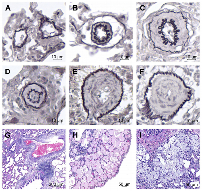
The monocrotaline (MCT)-induced pulmonary arterial hypertension model is one of the most reproduced today, presenting as a limitation the absence of plexiform lesions, typical manifestations of the severe disease in humans.
Objective
To evaluate the severity of MCT-induced pulmonary arteriopathy by pathological findings of lung and heart tissue samples, clinical course and 37-day survival.
Methods
Fifty male Wistar rats were divided into one of the four groups – control (CG) (n = 10) and three intervention (MCT) groups. The MCT groups received intraperitoneal injection (60 mg/kg) of MCT and remained exposed to the substance for 15 days (G15, n = 10), 30 days (G30, n = 10) and 37 days (G37, n = 20). At the end of each period, the animals were sacrificed, and pulmonary and cardiac tissues were collected for anatomopathological and morphometric analysis. The Kruskal-Wallis test was used, considering a level of significance of 5%.
Results
In the lungs of MCT animals, lesions related to pulmonary arteriopathy were found, including muscularization of the arterioles, hypertrophy of the middle layer and concentric neointimal lesions. Complex lesions were observed in MCT groups, described as plexiform and plexiform-like lesions. Right ventricular hypertrophy was evidenced by increased thickness and diameter of the cardiomyocytes and a significant increase in the right ventricular wall thickness (p <0.0000).
Conclusion
The MCT model was able to generate moderate-severe pulmonary arteriopathy associated with secondary right ventricular hypertrophy. The 37-day survival rate was 50%. To our knowledge, this study was the first to note the presence of complex vascular lesions, similar to those observed in patients with severe pulmonary arterial hypertension, in an isolated MCT model. (Arq Bras Cardiol. 2020; 115(3):480-490)
1,095