Arq. Bras. Cardiol. 2022; 119(6): 902-909
Right Ventricle Involvement by Glycogen Storage Cardiomyopathy (PRKAG2): Standard and Advanced Echocardiography Analyses
José Luiz Barros Pena
![]() , Fabricio Junqueira de Melo, Wander Costa Santos, Isabel Cristina Gomes Moura, Gabriela Pansanato Nakashima, Natalia Costa Freitas, Eduardo Back Sternick
, Fabricio Junqueira de Melo, Wander Costa Santos, Isabel Cristina Gomes Moura, Gabriela Pansanato Nakashima, Natalia Costa Freitas, Eduardo Back Sternick
This Original Article is referred by the Short Editorial "Focusing on the Right Ventricle in PRKAG2 Syndrome".
Abstract
Background
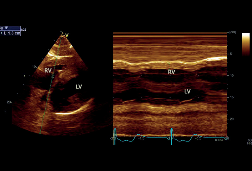
PRKAG2 syndrome is a rare, early-onset autosomal dominant inherited disease. We aimed to describe the right ventricle (RV) echocardiographic findings using two and three-dimensional (2D and 3D) modalities including myocardial deformation indices in this cardiomyopathy. We also aimed to demonstrate whether this technique could identify changes in RV function that could distinguish any particular findings.
Methods
Thirty patients with genetically proven PRKAG2 (R302Q and H401Q), 16 (53.3%) males, mean age 39.1 ± 15.4 years, underwent complete echocardiography examination. RV-focused, 4-chamber view was acquired for 2D and 3D measurements. Student’s t or Wilcoxon-Mann-Whitney tests were used to compare numerical variables between 2 groups, and p < 0.05 was considered significant.
Results
Twelve patients (40%) had a pacemaker implanted for 12.4 ± 9.9 years. RV free wall mean diastolic thickness was 7.9 ± 2.9 mm. RV 4-chamber longitudinal strain (RV4LS), including the free wall and interventricular septum, was –17.3% ± 6.7%, and RV free wall longitudinal strain (RVFWLS) was −19.1% ± 8.5%. The RVFWLS apical ratio measured 0.63 ± 0.15. Mean RV 3D ejection fraction (EF) was 42.6% ± 10.9% and below normal limits in 56.7% of patients. Positive correlation occurred between RV 3DEF, RV4LS, and RVFWLS, especially for patients without a pacemaker (p = 0.006).
Conclusion
RV involvement in PRKAG2 syndrome is frequent, occurring in different degrees. Echocardiography is a valuable tool in detecting RV myocardial abnormalities in this condition. The use of 2D RV4LS, RVFWLS, and 3DEF offers reliable indicators of RV systolic dysfunction in this rare, challenging cardiomyopathy.
571