Arq. Bras. Cardiol. 2021; 116(6): 1165-1168
Spiked Helmet Sign: An Atypical Case of Transient ST-Segment Elevation on ECG
Case report
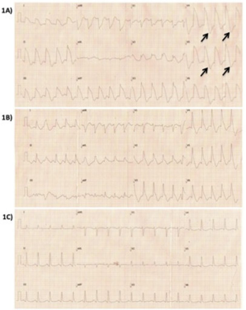
Male, 35 years old, smoker, user of illicit drugs (marijuana, cocaine and inhalant solvents), on antipsychotics and antidepressants (haloperidol and escitalopram), several episodes of vomiting and diarrhea for the past two days, mental confusion at home, and admitted to the emergency room with reduced level of consciousness and irregular breathing. Soon afterwards, he presented cardiopulmonary arrest (CRP) with ventricular fibrillation (VF) on the cardiac monitor. After six minutes of cardiopulmonary resuscitation maneuvers, adrenaline infusion and two cardiac defibrillations, pulse and heart rate were recovered. An electrocardiogram (ECG) was performed, which demonstrated atypical and diffuse ST-segment elevation ( ). Initial laboratory tests showed severe metabolic acidosis with high serum lactate (serum lactate = 26 mg/dl), in addition to hypernatremia (serum sodium = 153 mg/dl), hypokalemia (serum potassium = 3.2 mg/dl), severe leukocytosis and a slight increase in serum troponin. The patient was intubated and placed on mechanical ventilation. Volume resuscitation was performed, metabolic acidosis was corrected, and broad-spectrum antibiotics were initiated. One hour after initial consultation, a new ECG was performed, which demonstrated a reduction of approximately 50% in ST elevation ( ). Given the atypical character of the ST segment abnormalities on ECG, we chose not to perform an emergency coronary angiography. Six hours after the event, the initial ECG abnormalities receded completely ( ). An echocardiogram was performed, which showed severe left ventricular dysfunction at the expense of diffuse hypokinesia (ejection fraction = 0.36). A new echocardiogram was performed two days later, which revealed complete recovery of ventricular function (ejection fraction = 0.69). Coronary angiotomography was performed during hospitalization, which demonstrated the absence of obstructive lesions and ruled out coronary anomalies. The patient progressed well and was discharged after eight days of hospitalization.
[…]
1,390

