Arq. Bras. Cardiol. 2022; 118(6): 1099-1105
The Volume-Time Curve by Three-Dimensional Echocardiography in Chagas Cardiomyopathy: Insights into the Mechanism of Hemodynamic Adaptations
Airandes de Sousa Pinto
![]() , Maria Carmo Pereira Nunes
, Maria Carmo Pereira Nunes
![]() , Carlos Alberto Rodrigues
, Carlos Alberto Rodrigues
![]() , Bráulio Muzzi Ribeiro de Oliveira, João da Rocha Medrado Neto, Timothy C. Tan, Manoel Otavio da Costa Rocha
, Bráulio Muzzi Ribeiro de Oliveira, João da Rocha Medrado Neto, Timothy C. Tan, Manoel Otavio da Costa Rocha
![]()
This Original Article is referred by the Short Editorial "Is it Possible to Non-Invasively Study the Hemodynamic Adaptations of Chagas Cardiomyopathy by the Volume-Time Curve Using 3D Echocardiography?".
Abstract
Background
Three-dimensional echocardiography (3D ECHO) allows the generation of a volume-time curve representative of changes in the left ventricular (LV) volume throughout the entire cardiac cycle.
Objective
This study aims to demonstrate the hemodynamic adaptations present in Chagas cardiomyopathy (CC) by means of the volume and flow measurements obtained by the volume-time curve by 3D ECHO.
Methods
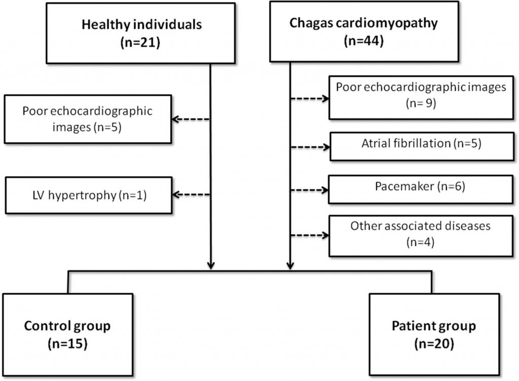
Twenty patients with CC and 15 healthy subjects were prospectively enrolled in a cross-sectional design study. 3D ECHO was performed in all subjects and the volume over time curves of the LV was generated. The flow was obtained by the first derivative of the volume-time curve using the software MATLAB. Statistical significance was set at p<0.05.
Results
Although CC patients had lower LV ejection fraction compared to the control group (29.8±7.5 vs. 57.7±6.1, p<0.001), stroke volume (61.5±25.2 vs. 53.8±21.0, p=0.364) and maximum ejection flow during systole (-360.3±147.5 vs. -305.6±126.0, p=0.231) were similar between the groups. Likewise, the maximum flow in the early diastolic filling phase and during atrial contraction was similar between groups. An increase in preload expressed by LV end diastolic volume (204.8±79.4 vs. 93.0±32.6), p<0.001) may maintain the flow and stroke volumes similar to the controls.
Conclusion
Using a non-invasive tool, we demonstrated that an increase in LV end-diastolic volume may be the main adaptation mechanism that maintains the flow and stroke volumes in the setting of severe LV systolic dysfunction.
842