Arq. Bras. Cardiol. 2021; 116(2 suppl 1): 36-38
Three-dimensional Echocardiography Reveals the True Enemy in a Young Male with ST-Elevation Myocardial Infarction and Severe Mitral Regurgitation: Posterior Mitral Valve “Pseudo-Cleft” and Prolapse
Sorina Mihaila
![]() , Andreea Elena Velcea, Luigi Paolo Badano, Vinereanu Dragos, Denisa Muraru
, Andreea Elena Velcea, Luigi Paolo Badano, Vinereanu Dragos, Denisa Muraru
Introduction
Three-dimensional echocardiography (3DE) plays an increasingly important role in the diagnosis of valvular heart disease, in the assessment of valvular morphology in an anatomical manner, and in establishing valve repairability, beyond the limitations of conventional two-dimensional echocardiography (2DE).
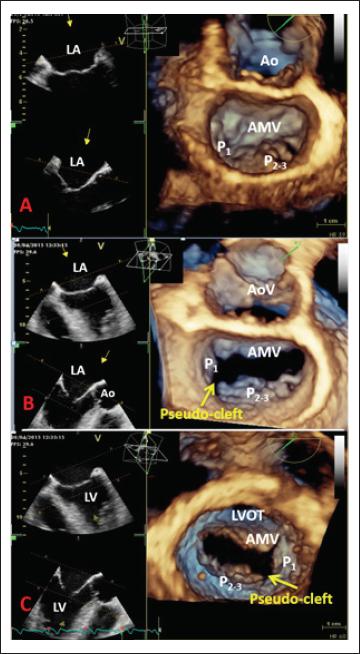
We report the case of a young patient presenting with acute anterior ST-segment elevation myocardial infarction and severe mitral regurgitation (MR) after successful primary percutaneous coronary intervention (PCI) of the left anterior descending artery, whose three-dimensional transesophageal echocardiography (3D TEE) revealed an unexpected cause of the MR, namely, complex mitral valve (MV) pathology consisting of prolapse of the P2-3 scallops, flail chordae, and pseudo-cleft of the posterior leaflet separating the P1 from the P2 segment.
[…]
1,248