Arq. Bras. Cardiol. 2022; 119(3): 499-501
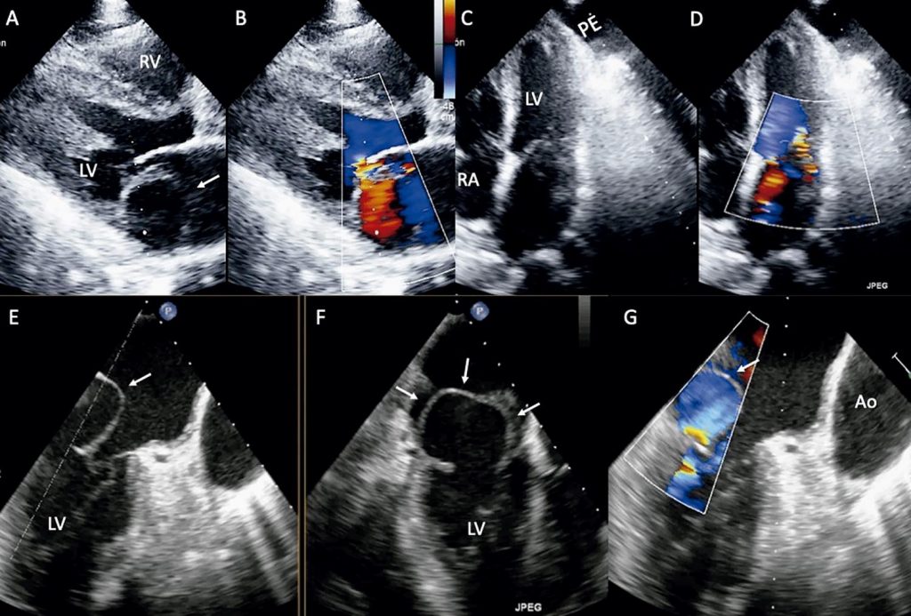
Transesophageal Two- and Three-Dimensional Echocardiographic Assessment of Spontaneous Left Atrial Dissection
Javier Ivan Armenta-Moreno
![]() , Joaquin Berarducci, Abel Mauricio Garcia-Cardenas, José Carlos Armendariz-Ferrari, Jorge Luis Bermudez-Gonzalez, Juan Ignacio Straface, Jose Antonio Luna-Alvarez-Amezquita, Nilda Espinola-Zavaleta
, Joaquin Berarducci, Abel Mauricio Garcia-Cardenas, José Carlos Armendariz-Ferrari, Jorge Luis Bermudez-Gonzalez, Juan Ignacio Straface, Jose Antonio Luna-Alvarez-Amezquita, Nilda Espinola-Zavaleta
![]()
A 41-year-old woman was admitted to the emergency room with an onset of acute dyspnea, jugular ingurgitation, and with the first heart sound diminished, followed by a grade III/IV holosystolic murmur that was better heard at the apex and edema of the lower limbs.
The vital signs were heart rate 91 bpm, respiratory rate 21 rpm, blood pressure 110/60 mmHg, and oxygen saturation 91%. The chest x-ray showed cardiomegaly with a cardiothoracic index of 0.62 and pulmonary venocapillary hypertension.
[…]
322