Arq. Bras. Cardiol. 2023; 120(3): e20220573
Tricky Diagnosis: An Aberrant Mitral Valve Chord
Marina Santos
![]() , Mariana Paiva, Joana Ferreira
, Mariana Paiva, Joana Ferreira
![]() , Sara Guerreiro
, Sara Guerreiro
![]()
Case
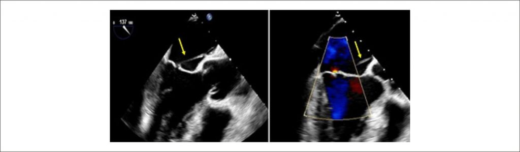
We present the case of a 52-year-old man with hepatitis C and a history of intravenous drug abuse, admitted to the infectology department with spondylodiscitis and psoas muscle abscess. Staphylococcus aureus was isolated from blood, urine, and spinal fluid cultures, and the transthoracic echocardiogram was negative for vegetation, abscess, or fistula. The transesophageal echocardiogram (TEE) showed a filamentous and mobile structure attached to the atrial surface of the mitral valve (MV), and a diagnosis of infective endocarditis was presumed. Seven days later, the patient was referred to our tertiary institution for a detailed TEE. In 2D images, we found a thin, well-delineated strand connecting the interatrial septum (IAS) to the tip of the anterior mitral leaflet without significant regurgitation ( , supplementary video 1). 3D imaging confirmed the presence of an anomalous chord connecting the A2 scallop of MV to the middle of the interatrial septum ( , supplementary video 2). There was no evidence of infective endocarditis, and this strand was found to be compatible with an anomalous left atrial MV chord.
[…]
635