Arq. Bras. Cardiol. 2021; 117(4): 688-689
Myocardial Scar and Ventricular Repolarization on the electrocardiogram: Insights from Cardiac Magnetic Resonance Imaging
This Short Editorial is referred by the Research article "Relationship between Late Gadolinium Enhancement and Ventricular Repolarization Parameters in Heart Failure Patients with Reduced Ejection Fraction".
Myocardial fibrosis is a final common feature of most inflammatory pathway and is frequently seen in patients with heart failure with reduced ejection fraction (HFrEF). , Myocardial scar, detected by contrast enhanced magnetic resonance imaging (ce-MRI), has been shown to be a predictor of prognosis and sudden cardiac death in patients with HFrEF. – Cardiac MRI has emerged as a non-invasive gold standard for the assessment of scar and risk prediction of ventricular arrhythmias. , , , Nevertheless, cardiac MRI can be time consuming, expensive, and not easily available.
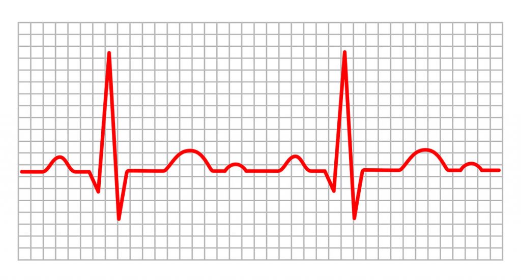
The 12-lead surface electrocardiogram (ECG) is a simple and inexpensive diagnostic tool that is widely used in clinical practice for the diagnosis of cardiac arrhythmias. The ECG waveform is the expression of the transmembrane action potentials and is composed of two distinct processes: depolarization and repolarization. Ventricular repolarization is a complex electrical phenomenon which in clinical practice is measured from the beginning of the QRS complex to the end of the T-wave. Cardiac structural and electrical changes may cause action potential abnormalities, affecting the refractory period and thus favoring the onset of ventricular arrhythmias.
[…]
3,044

